
Why Imaging Is Often Ordered Before Symptoms Become Severe
Medical imaging helps physicians see structures inside the body that cannot be fully evaluated through symptoms, conversation, or physical examination alone. It can show bones, organs, blood vessels, soft tissues, breast tissue, the brain, joints, the abdomen, the chest, or other areas depending on the test. Imaging does not replace clinical judgment, but it can give the care team important visual information when a symptom, screening need, abnormal exam finding, or previous test result requires clarification.
Many patients think imaging is only used after pain becomes severe or when a disease is already obvious. In reality, imaging can also be used earlier in care when a physician needs to investigate risk, screen selected patients, monitor a known condition, or better understand a finding before symptoms become advanced. For example, mammography may be used to screen eligible women for breast cancer before symptoms appear, while low-dose CT is the recommended screening test for lung cancer in selected higher-risk adults. These tests are used for specific populations and specific clinical questions, not as general scans for everyone.
Imaging can also help when symptoms are vague. Abdominal discomfort, persistent headaches, joint pain, swelling, shortness of breath, pelvic symptoms, breast changes, injury, unexplained weight loss, or abnormal laboratory results may lead a physician to order a specific imaging study. The choice depends on what the clinician needs to see. A CT scan, MRI, ultrasound, X-ray, and mammogram are not interchangeable tests.
The value of imaging is not that it “finds everything,” but that it can answer a focused medical question when the right test is matched to the patient’s symptoms, risk factors, exam findings, and health history.
RadiologyInfo, a patient resource from the American College of Radiology and the Radiological Society of North America, explains that radiology information is used to describe how X-ray, CT, MRI, ultrasound, radiation therapy, and other procedures are performed, what patients may experience, and how to prepare. That patient-facing focus matters because many people arrive for imaging without understanding why the test was chosen or what the result can and cannot show.
A physician may also order imaging after a laboratory result. A blood test may suggest inflammation, liver or kidney involvement, infection, anemia, or another concern, but lab work cannot always show where the problem is located or what structure is involved. Imaging may help connect laboratory data with anatomy. Patients who want to understand that pathway can review laboratory results that help physicians make faster decisions.
Routine care can also lead to imaging when appropriate. During a preventive visit, a physician may identify that a patient is due for mammography, has a symptom that needs ultrasound, has a smoking history that requires a lung screening discussion, or has pain that deserves further evaluation. Patients who want to understand where imaging fits into prevention can review preventive visits that help determine when testing is needed.
However, imaging should not be ordered casually. Some tests involve radiation, contrast material, incidental findings, cost, anxiety, and follow-up procedures. The FDA notes that CT scans carry risks such as incidental results that may lead to unnecessary follow-up testing, while the CDC explains that mammograms use a small amount of radiation and may also miss some cancers or lead to additional testing. This is why imaging decisions should balance benefit and risk.
MRI, CT, Ultrasound and Mammography: What Each Test Is Used For
A Sweetwater medical center may offer several types of exams, but each one works differently. The best test depends on the clinical question. Patients should not assume that the most expensive test is automatically the best, or that one scan can replace all others. A physician chooses imaging based on what body area needs evaluation, how urgent the concern is, what level of detail is needed, whether radiation should be avoided, whether contrast is required, and whether the patient has medical limitations such as implants, kidney disease, pregnancy, allergy history, or claustrophobia.
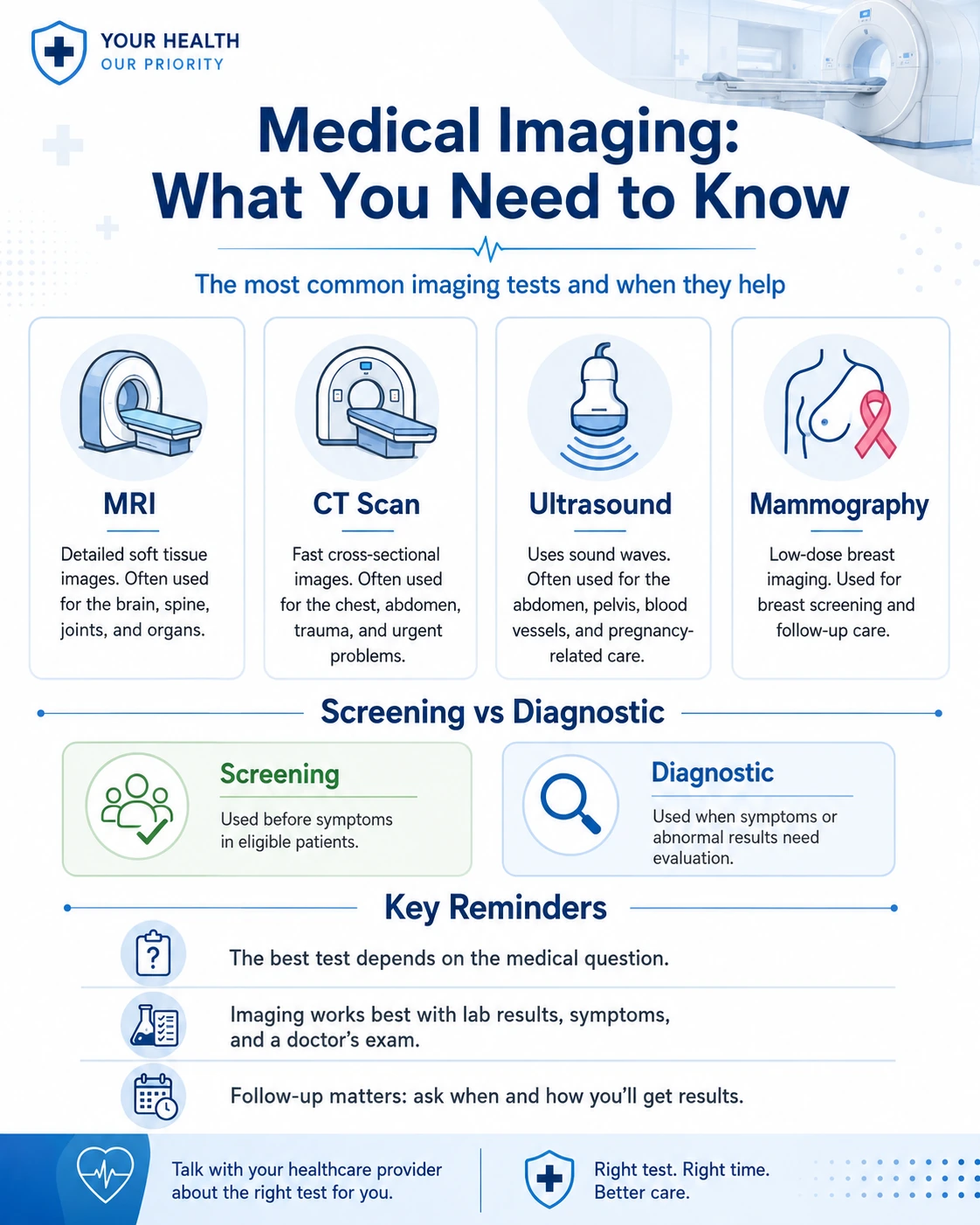
An MRI scan uses a strong magnetic field and radiofrequency energy to create detailed images of the body. MRI can be useful for many soft tissue, brain, spine, joint, pelvic, abdominal, vascular, and neurologic questions depending on the protocol. It does not use ionizing radiation, but it is not appropriate for every patient. Certain implants, metal fragments, devices, severe claustrophobia, or inability to lie still may affect whether MRI is safe or practical.
A CT scan uses X-rays and computer processing to create cross-sectional images. CT can be fast and detailed, which makes it useful in many emergency, chest, abdominal, trauma, lung, vascular, and cancer-related evaluations depending on the situation. CT does use ionizing radiation, so the decision should be based on medical need. In some cases, contrast material is used to improve detail, and the care team may need to review kidney function, allergy history, or other risk factors before the exam.
Ultrasound imaging uses sound waves to create real-time images. It is commonly used for abdominal, pelvic, vascular, breast, thyroid, pregnancy-related, soft tissue, and procedure-guidance questions depending on the indication. Ultrasound does not use ionizing radiation and can show movement or blood flow in selected studies. Its usefulness can depend on the body area, patient anatomy, bowel gas, operator technique, and the clinical question being asked.
Mammography uses low-dose X-rays to create images of breast tissue. It may be used as a screening test in eligible women or as part of diagnostic evaluation when a breast symptom or abnormal screening result needs follow-up. The CDC explains that mammograms use X-ray beams to create images of the inside of the breast and can help diagnose breast cancer and other conditions. Patients should understand whether their appointment is for screening or diagnostic imaging because the purpose and follow-up process may differ.
Other imaging methods may also be used depending on the medical center, including plain X-ray, fluoroscopy, nuclear medicine, PET/CT, bone density testing, and image-guided procedures. This article focuses on the most common modalities patients ask about, but the same principle applies to all imaging: the test should match the question.
Imaging Methods and Typical Clinical Uses
The table below gives a general overview of how common imaging methods may be used. It is not a personal recommendation. The right test depends on the physician’s order, symptoms, risk factors, preparation requirements, and safety considerations.
| Method | What it can show | Common reasons it may be ordered | Patient experience |
|---|---|---|---|
| Magnetic resonance exam | Detailed soft tissue, brain, spine, joint or organ information depending on protocol | Neurologic, musculoskeletal, abdominal, pelvic or vascular questions | The patient lies still inside a scanner; noise and enclosed space may be noticeable |
| Computed tomography | Cross-sectional views of internal structures | Trauma, chest, abdomen, lung, vascular, infection or cancer-related evaluation | The scan is usually fast; contrast may be used in selected cases |
| Sonographic exam | Real-time images of organs, vessels, soft tissue or fluid | Abdominal, pelvic, thyroid, breast, vascular or pregnancy-related questions | A probe is moved over the skin with gel; the exam is usually not painful |
| Breast X-ray exam | Internal breast tissue patterns | Routine breast screening or follow-up of a breast concern | Compression is used briefly to improve image quality |
| Plain radiograph | Bones, chest structures and selected abdominal findings | Fracture, chest infection, joint, spine or injury evaluation | The exam is usually quick and may require positioning |
| Bone density exam | Bone mineral density | Fracture risk or osteoporosis evaluation in selected patients | The exam is usually brief and noninvasive |
Screening vs Diagnostic Imaging: Why the Difference Matters
One of the most important distinctions in radiology is the difference between screening and diagnostic imaging. Screening is used in selected people who meet criteria before symptoms appear. Diagnostic imaging is used when there is a symptom, abnormal physical exam, abnormal screening result, abnormal laboratory finding, injury, or other medical reason to investigate a specific concern.
For example, a screening mammogram is performed for eligible women without breast symptoms. A diagnostic breast exam is used when there is a lump, nipple discharge, skin change, focal pain, or an abnormal screening result. These two exams may feel similar to the patient, but they answer different clinical questions and may have different imaging views, timing, interpretation, and follow-up.
Lung cancer screening is another example. The CDC states that the only recommended screening test for lung cancer is low-dose CT. This is not a general chest scan for anyone who is worried. It is intended for eligible higher-risk adults based on age and smoking history. If a patient has symptoms such as coughing blood, persistent chest pain, unexplained weight loss, or worsening shortness of breath, the question becomes diagnostic, not routine screening.
Screening imaging can find disease before symptoms, but it can also find abnormalities that are not cancer or not dangerous. These incidental or unclear findings may require follow-up imaging, comparison with previous studies, biopsy, or specialist review. That does not mean screening was wrong. It means patients should understand the possible next steps before testing begins.
Diagnostic imaging can also be normal even when symptoms are real. A normal scan may reduce concern for certain conditions, but it does not always explain pain, fatigue, dizziness, or other symptoms. The physician may need lab work, medication review, physical therapy, specialist evaluation, or follow-up if symptoms continue.
Patients reviewing broader prevention can use cancer screening recommendations by age to understand when screening is appropriate. Women planning preventive care can also review a women’s screening checklist that connects mammography, cervical health, bone health, and cardiovascular risk.
When Imaging Follows a Lab Result or Physical Exam
Imaging is often ordered after a physician has already gathered other information. A symptom, physical exam finding, laboratory result, screening result, or previous medical history may point to a question that cannot be answered fully without looking inside the body. This is why imaging and laboratory testing often work together rather than compete with each other.
For example, abdominal pain may begin with a history, physical exam, and laboratory testing. If the physician is concerned about inflammation, infection, gallbladder disease, kidney stones, liver or pancreas involvement, bowel problems, or another structural issue, imaging may be ordered to clarify the location and possible cause. The exact test depends on the suspected condition and urgency.
Abnormal liver markers may lead to ultrasound, CT, MRI, or other evaluation depending on the pattern and symptoms. Blood in the urine may lead to imaging of the urinary tract in selected cases. Anemia may lead to additional laboratory work, endoscopy, imaging, or other evaluation depending on age, symptoms, severity, and history. A physical exam finding such as a mass, swelling, tenderness, or abnormal breast finding may also lead to targeted imaging.
In injury care, the physical exam often guides the imaging choice. A minor sprain may not need imaging, while severe pain, deformity, inability to bear weight, neurologic symptoms, or high-impact trauma may require X-ray, CT, MRI, or ultrasound depending on the suspected injury. The goal is to evaluate what is clinically relevant, not to image every painful area automatically.
In cancer screening and follow-up, imaging may follow an abnormal screening result. An abnormal mammogram may lead to diagnostic mammography, ultrasound, MRI in selected cases, or biopsy. A lung screening finding may lead to repeat CT, specialist review, PET imaging, biopsy, or surveillance depending on size, appearance, and risk. A screening test is only useful when the next step is clear.
Laboratory and imaging results also need physician interpretation. A scan may show a finding that is incidental, age-related, benign, uncertain, or clinically important. A lab result may be abnormal but temporary, medication-related, or expected in the patient’s condition. The physician’s role is to combine the pieces and decide what matters now.
Patients should ask why imaging is being ordered and what question it is meant to answer. Useful questions include: what condition is being considered, whether contrast is needed, whether previous images should be compared, what the possible next steps are, and how results will be communicated. Clear expectations reduce anxiety and help patients understand the purpose of testing.
What Patients Should Know Before an Imaging Appointment
Preparation depends on the type of exam. Some imaging tests require no special preparation. Others may require fasting, drinking water, avoiding certain medications, checking kidney function, removing metal objects, bringing previous images, or arriving early for contrast screening. Patients should follow the instructions from the imaging center rather than relying on general advice.
Safety questions are important. Before MRI, patients may be asked about pacemakers, implanted devices, metal fragments, aneurysm clips, cochlear implants, medication pumps, surgical hardware, or prior injuries involving metal. Before CT or MRI with contrast, the team may ask about kidney function, allergy history, pregnancy status when relevant, breastfeeding, diabetes medications, or previous contrast reactions. These questions are routine safety steps, not signs that something is wrong.
Patients should also bring relevant records when possible. Prior imaging reports, image discs or digital access, previous biopsy results, operative notes, lab results, and specialist records can help the radiologist compare findings over time. Comparison can be especially important for lung nodules, breast findings, cysts, masses, spine changes, and chronic conditions.
Comfort concerns should be discussed before the exam. Claustrophobia, pain when lying flat, mobility limitations, difficulty holding still, hearing sensitivity, anxiety, or need for assistance may affect how the appointment is planned. Some facilities can offer positioning support, communication tools, open or wide-bore scanners where available, music, breaks, or other accommodations depending on the exam.
Before Your Imaging Visit
A short preparation checklist can help patients avoid delays and incomplete exams:
- Follow the preparation instructions from the imaging center
- Bring the physician order or referral if required
- Tell the team about implants, devices or metal fragments
- Ask whether contrast will be used and whether lab work is needed first
- Bring prior imaging reports or access information when available
- Mention pregnancy status when relevant
- Wear comfortable clothing and remove metal objects when instructed
- Ask when and how results will be sent to the ordering clinician
Patients should not assume they will receive a final explanation from the technologist during the exam. Technologists are trained to perform the study and obtain high-quality images, but the final interpretation usually comes from a radiologist. The ordering physician then connects the imaging report with symptoms, exam findings, labs, and the patient’s history.
How Imaging Results Are Interpreted
After the exam, images are reviewed by a radiologist or another qualified specialist depending on the setting and type of study. The radiologist prepares a report for the ordering clinician. This report may describe what was seen, what was not seen, whether findings are new or stable, whether comparison with previous images was available, and what follow-up may be considered.
The report may include technical language that is difficult for patients to understand. Words such as lesion, nodule, density, opacity, cyst, degenerative change, enhancement, calcification, artifact, or incidental finding do not all mean the same thing and do not automatically mean a dangerous disease is present. The meaning depends on body area, size, appearance, symptoms, history, risk factors, and comparison with prior studies.
In many cases, the ordering physician is the best person to explain what the result means for the patient. A radiology report answers an imaging question, but the treating clinician knows why the test was ordered and how the result fits with the patient’s condition. For example, a spine MRI may show age-related changes in a patient whose pain is actually coming from another source. A CT may show a small incidental finding that needs no urgent treatment but should be documented. A mammogram may require additional views even though the final result may be benign.
Patients should ask whether the result changes the plan. A useful explanation should include whether the finding is normal, abnormal but not urgent, uncertain, needs follow-up, needs specialist referral, or requires immediate care. The report should also lead to a clear next step: observation, repeat imaging, lab testing, medication, procedure, biopsy, physical therapy, surgery consultation, or reassurance when appropriate.
Results can also be affected by image quality and patient factors. Movement, body position, bowel gas, metal artifacts, incomplete preparation, timing of contrast, or inability to complete the exam can limit what the radiologist can see. If a report says the study is limited, the physician may decide whether another test or repeat study is needed.
When imaging happens during a hospital stay, emergency visit, or complex medical evaluation, several professionals may be involved. The technologist performs the exam, the radiologist interprets the images, nurses and physicians monitor patient safety, laboratory staff may process related tests, and the treating physician decides the care plan. Patients interested in this broader team structure can review the care team involved in your hospital stay.
Common Myths About Imaging
Medical imaging is powerful, but misunderstandings can lead to anxiety, unnecessary testing, or false reassurance. Patients often hear about scans from friends, online forums, advertisements, or personal stories. Those experiences may not apply to their own symptoms, risks, or medical history.
Myth: More Imaging Is Always Better
More imaging does not always mean better care. Unnecessary imaging can expose patients to radiation in some tests, lead to incidental findings, increase cost, and trigger follow-up procedures that may not improve health. The right question is not “Can we scan it?” but “Will this test help answer a clinically useful question?”
Myth: A Normal Image Means Nothing Is Wrong
A normal imaging result can be reassuring, but it does not explain every symptom. Pain, fatigue, dizziness, weakness, digestive symptoms, or shortness of breath may require lab work, medication review, functional assessment, sleep evaluation, specialist care, or follow-up. A scan is one part of the evaluation.
Myth: All Scans Use the Same Technology
MRI, CT, ultrasound, X-ray, mammography, and bone density testing use different technologies and answer different questions. A test that is excellent for one condition may be less useful for another. This is why physicians choose imaging based on the clinical question rather than patient preference alone.
Myth: Imaging Replaces the Doctor’s Exam
Imaging can show anatomy, but it does not replace the medical history, physical exam, laboratory results, medication review, and clinical judgment. The same image finding may be important in one patient and incidental in another.
Myth: Every Finding Is Dangerous
Imaging can reveal benign, age-related, old, stable, or incidental findings. Some need follow-up, while others do not. Patients should avoid assuming the worst before the ordering physician explains the result.
How to Prepare for Results and Follow-Up
Many patients focus on the scan itself but do not think about what happens after the images are taken. Follow-up is part of the imaging process. A study is not complete until the result is reviewed, explained, and connected to a clear next step. Without that step, patients may misunderstand findings, miss important follow-up, or become unnecessarily anxious about medical language in the report.
Before leaving the appointment, patients should know who will receive the report, whether results will appear in a portal, how long interpretation may take, and whether the ordering physician will contact them directly. Some findings require rapid communication, while others are reviewed during a scheduled follow-up. The timing depends on the urgency of the clinical question and the facility’s process.
If the report recommends comparison with prior images, patients may need to help obtain those records. Prior studies can show whether a finding is new, stable, growing, shrinking, or previously known. This can change the meaning of the result. A small finding that has been stable for years may be handled differently from a new finding with concerning features.
Patients should also ask what symptoms should prompt earlier contact. If pain worsens, fever develops, breathing changes, neurologic symptoms appear, bleeding occurs, or the original concern becomes severe, waiting for a routine result review may not be appropriate. Imaging follow-up should not delay urgent care when symptoms change.
When additional testing is recommended, patients should ask why. A follow-up scan, biopsy, lab test, specialist visit, or procedure may be ordered because the first test found something that needs clarification. This does not automatically mean a serious diagnosis. It means the care team needs more information before making a confident decision.
How Imaging Supports Earlier and Safer Care
Imaging can support earlier care when it is used for the right patient and the right reason. It may help physicians detect selected diseases before symptoms become obvious, clarify symptoms before complications develop, or monitor known findings over time. This is especially important in breast screening, lung screening for eligible higher-risk adults, injury evaluation, vascular concerns, abdominal symptoms, pelvic symptoms, and follow-up after abnormal tests.
However, earlier does not always mean better if the test is not appropriate. An unnecessary scan can create incidental findings, repeat testing, radiation exposure in some cases, and anxiety without improving care. A physician’s role is to decide whether imaging is likely to change management. If the result will not affect treatment, follow-up, or risk assessment, the test may not be useful.
Imaging also helps avoid delay when symptoms are concerning. For example, a patient with severe abdominal pain, neurologic symptoms, suspected fracture, abnormal breast finding, or complicated infection may need imaging to decide the next step quickly. In these situations, imaging is not a general wellness tool; it is part of urgent or diagnostic decision-making.
For preventive care, imaging should fit into a larger plan. A screening mammogram, bone density exam, or low-dose lung CT should not be ordered in isolation without a clear process for results and follow-up. Patients should know why the test is recommended, what an abnormal result may require, and when the next screening should be discussed.
The phrase mammography is often associated with routine breast screening, but it is also a reminder that imaging works best when patients understand the purpose of the exam. A screening mammogram, diagnostic mammogram, ultrasound, MRI, CT, or X-ray can all be valuable when matched to the correct clinical question. The test itself is only one part of care; interpretation and follow-up complete the process.
Medical Disclaimer
This article is for educational purposes only and does not replace professional medical advice, diagnosis, or treatment. Imaging decisions should be made with a qualified clinician based on symptoms, risk factors, medical history, examination findings, prior results, and current guidelines.
Author
By Dr. Cody R. Christensen, M.D. He practices at Sweetwater Medical Center, where he integrates pharmacologic treatment with lifestyle medicine and psychotherapy to support lasting patient wellness.
Medically Reviewed: by Clinical Pharmacy Board
Last Updated: 08.06.2026
FAQ
Can imaging detect disease before symptoms?
Yes, selected imaging tests can help detect certain conditions before symptoms appear when used for the right patient group. Examples include screening mammography for eligible women and low-dose CT for selected higher-risk adults being screened for lung cancer.
Which is better: MRI, CT or ultrasound?
No single test is best for every situation. The right choice depends on the body area, symptoms, urgency, safety factors, and the medical question your physician needs to answer.
Do I need a doctor’s order?
Many imaging exams require a clinician’s order because the test must match a specific medical reason. The ordering clinician also receives the report and connects the findings to your care plan.
Are imaging tests painful?
Most imaging tests are not painful, though some may involve pressure, positioning, an injection, contrast, or lying still for a period of time. Tell the imaging team if you have pain, anxiety, mobility limits, or trouble staying still.
What happens after the report is ready?
The radiologist sends a report to the ordering clinician, who explains what the findings mean in context. The next step may be no action, follow-up imaging, lab work, medication, referral, biopsy, or urgent care depending on the result.